Uterus transplantation is a medical frontier that defies conventional expectations and raises questions about the lengths certain individuals are willing to go to fulfill their dreams of parenthood.
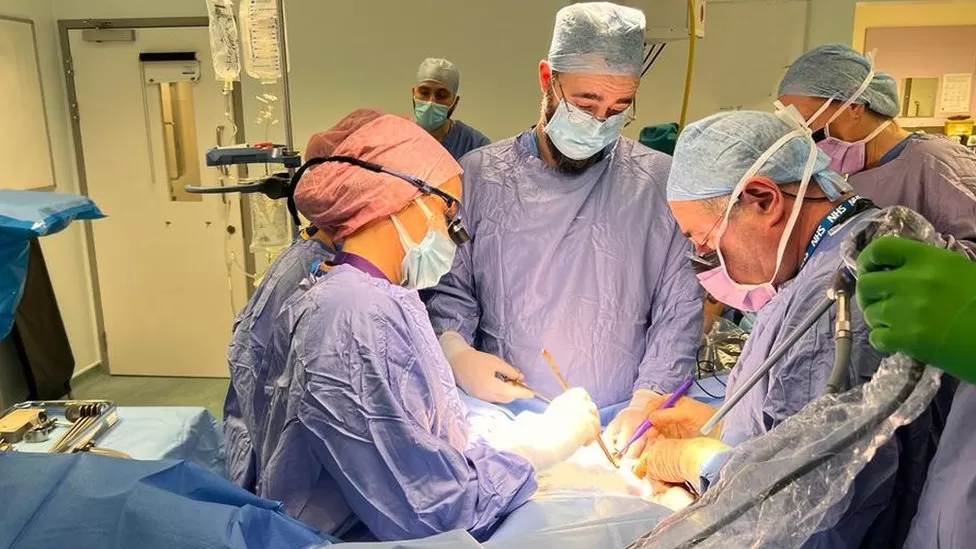
On August 23, a milestone was achieved at the Churchill Hospital Oxford in the United Kingdom, where the country’s first uterus transplant was performed.
In this groundbreaking procedure, the uterus of a 40-year-old woman was transplanted into her 34-year-old sister, who had a rare condition affecting her reproductive abilities, as reported by the BBC.
While the surgery was successful, the ultimate proof of its success lies in the hope of a live birth in the near future, according to Isabel Quiroga, the lead surgeon involved in the procedure.
Unlocking Uterus Transplants
Uterus transplants, unlike life-saving organ transplants, offer the promise of improving the quality of life for women who lack a uterus due to congenital conditions or other factors. These transplants can open the doors to fulfilling their (and possibly biological men) reproductive desires.
The first successful uterus transplant that led to a live birth occurred in Sweden in 2014. Dr. Mats Brännström, the chair of the obstetrics and gynecology department at the University of Gothenburg, led this groundbreaking initiative.
The success of this procedure signaled a potential solution for uterine factor infertility, with nine women in Sweden receiving transplanted wombs donated by relatives as part of Dr. Brännström’s program.

The challenge now is to make uterus transplants more accessible and affordable. In the United Kingdom, the National Health Service estimates the cost of the surgery at approximately GBP 25,000. In contrast, India has successfully performed uterus transplants, making this procedure more accessible, with the cost ranging from Rs 15-17 lakh.
The Journey of Uterus Transplantation
Uterus transplantation involves a series of critical steps.
First, the recipient undergoes a thorough evaluation for physical and mental health. The donated uterus, whether from a deceased or a living donor, is rigorously examined for viability.
Live donors also undergo extensive gynecological examinations, including CT and MRI scans, uterine cancer screening, and testing for the human papillomavirus.
Unlike the natural reproductive process, the procedure does not connect the transplanted uterus to the recipient’s fallopian tubes.
Instead, embryos are created through in vitro fertilization, and these embryos are cryopreserved. After the transplanted uterus is deemed ready, the embryos are implanted, allowing the recipient to pursue pregnancy.
During the transplantation, the uterus is carefully removed from the donor, a process now made less invasive with the assistance of robot-assisted laparoscopy.
The uterine transplant includes the removal of the arterial and venous vasculature, which consists of the deep uterine artery, the internal iliac arteries, the deep uterine vein, and the internal iliac veins.
Additionally, the fallopian tubes are removed and not used in the graft to prevent pregnancies outside the uterus. Surgical teams meticulously connect muscles, cartilage, tendons, and the various blood vessels to ensure that the transplanted uterus functions normally.
In a remarkable development, a surgeon in India has expressed interest in attempting a uterus transplant for a trans woman with the goal of enabling pregnancy, the Daily Mail reported.
This procedure would involve utilizing reproductive organs from a deceased donor or from a patient who has transitioned in the opposite direction. Such a procedure poses significant challenges and has been attempted only once in the past, with tragic results.
It would require in vitro fertilization and a C-section, as trans women do not possess fully functional vaginas.
Dr. Narendra Kaushik, who runs a gender reassignment clinic in New Delhi, is optimistic about the procedure, stating, “Every transgender woman wants to be as female as possible — and that includes being a mother.”
Pregnancy After Uterus Transplant
Surgeons assess the success of a uterus transplant in three stages. The first three months post-transplant are crucial, as this period carries the highest risk of graft loss.
After six months to a year, doctors monitor the proper functioning of the uterus, with regular menstruation signaling positive progress. Only after this phase can the recipient attempt conception.
The initial stages of pregnancy involve the transfer of embryos prepared through in vitro fertilization and cryopreserved.
However, pregnancies following uterine transplants carry certain risks, including potential graft rejection, spontaneous abortion, intrauterine death, low birth weight, and premature birth.
Consequently, frequent medical check-ups and follow-ups are imperative for women with transplanted uteri.
Understanding the Side Effects
To prevent the recipient’s body from rejecting the transplanted uterus, immunosuppressive drugs are necessary.
These drugs are carefully selected to ensure they do not harm fetal development during pregnancy. While immunosuppressants are vital, they do come with potential side effects, including kidney toxicity, bone marrow toxicity, a heightened risk of diabetes, and cancer.
For this reason, the uterus is typically removed after childbirth. Recipients are advised to undergo regular follow-up appointments for at least a decade following the uterus’s removal to monitor long-term side effects of immunosuppressants.
The Future: Artificial Uteri
Successful uterus transplants have paved the way for potential breakthroughs, such as transplanting uteri from deceased donors, an approach that has seen limited success.
This method could help alleviate the ethical and social concerns associated with using live donors, who undergo significant medical procedures to benefit another individual.
Dr. Mats Brännström and his team are also working on developing bioengineered artificial uteri. Such advancements could eliminate the need for live donors and address ethical debates surrounding organ transplantation.
The creation of a bioengineered uterus begins with a small cluster of stem cells extracted from a woman’s blood or bone marrow, serving as the foundation for a 3D scaffold. Additional cells are added to this scaffold to construct a functional uterus.
Although these experiments are in the early stages, initial results with animal testing have shown promise. However, it is estimated that it will take about a decade before artificial uteri reach the efficiency and safety required for human use.
This advancement could potentially benefit not only women but also members of the LGBTQ+ community.
Trans women, for example, may still need to undergo castration to create an artificial vagina. This process involves hormones, which can pose a threat to pregnancy, necessitating the administration of counteracting exogenous hormones.
Nevertheless, challenges remain in ensuring consistent blood flow to support fetal development, as the male body lacks the necessary structural elements for both a uterus and fetal development.
